

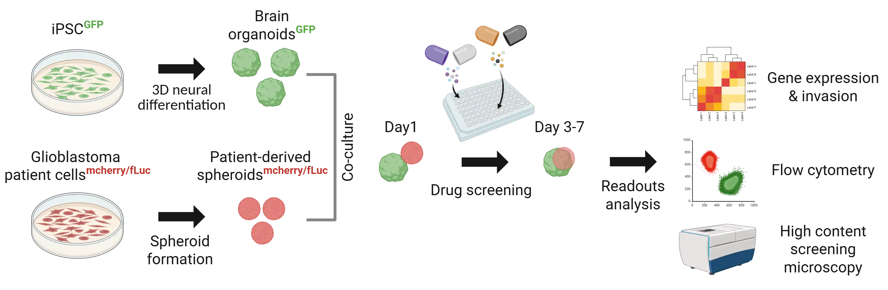
Our drug testing services allow us to evaluate compounds against brain tumours in a relevant neural environment. We create tumour spheroids from human glioblastoma cells that are either patient-derived or cell line-based.
Fluorescent markers are used to engineer these cells before inoculating them onto healthy brain organoid. Our approach assesses the therapeutic window of drugs while analysing their dual impact on tumour invasion and gene expression in both tumour and healthy tissues, providing comprehensive insights.
Unique features

Human 3-dimensional tissues
Short & long-term compound testing
Captures critical tumour–organoid interactions
01.
Patient-derived glioblastoma spheroid biobank faithfully reflecting the complexity and heterogeneity of tumour invasion and therapeutic response
02.
Co-culture with brain organoids for dual impact assessment
03.
Highly standardized glioblastoma cell line models as internal reference
Scaffold-free spheroid generation
Brain organoid differentiation
Patient-derived spheroid establishment
Assembloid generation (healthy and tumour)
2D/3D imaging
Immunohistochemistry / Immunofluorescence
Fluorescence-based assays
Flow cytometry
Genomics, Proteomics
Quantification of tumour invasiveness into the healthy brain organoid.
Highly reproducible human model
Reproduces patient heterogeneity in proliferation and invasive states

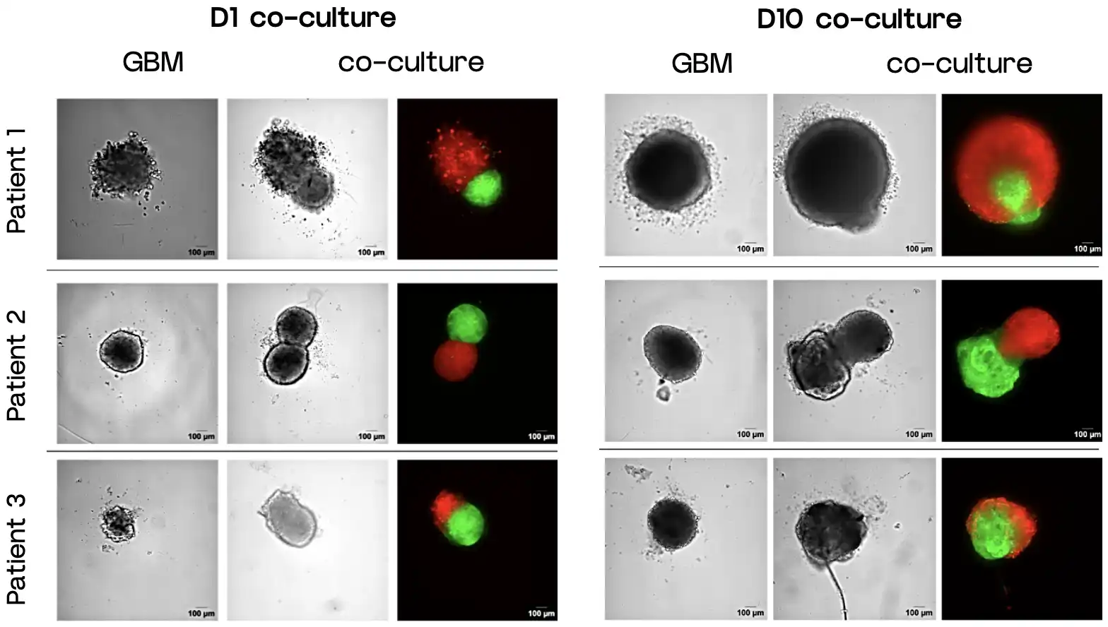
Representative brightfield and confocal microscope images of patient-derived glioblastoma in red (GBMmcherry/fLuc) co-cultured with brain-organoid (Neuro GFP) in green (NeuroGFP).
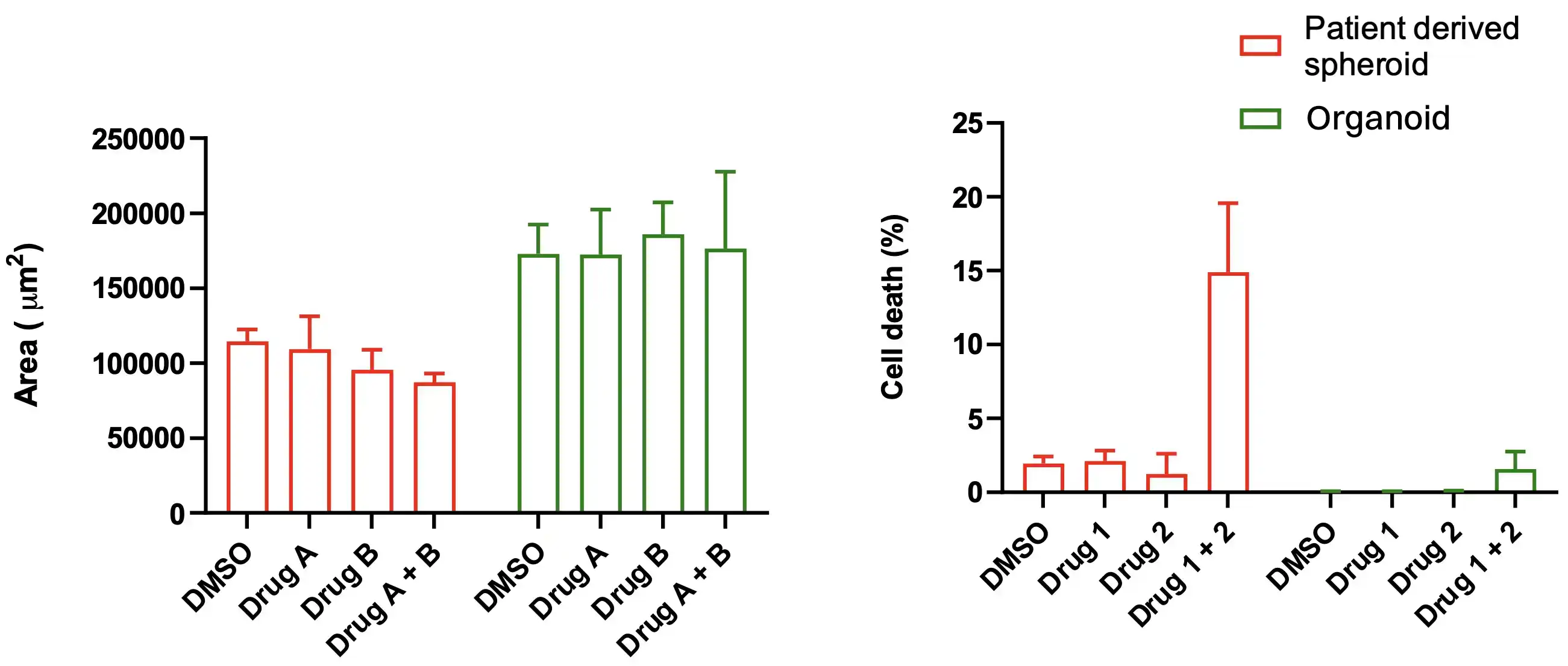
Quantitative analysis of the tumour area and viability upon drug treatment, measured by microscopy and flow cytometry after Draq7 staining, respectively.
Glioblastoma-organoid assembloids are a relevant model to assess drug efficiency on tumour growth and invasive states, while also testing effects on healthy tissue.
