BLASTOMABRAINTM GLIOBLASTOMA INVASION MODEL
GLIOBLASTOMA INVASION MODEL
HUMAN BRAIN-TUMOR ORGANOID MODEL
ADVANTAGES OF BLASTOMABRAINTM GLIOBLASTOMA INVASION MODEL
- Short-term and long-term compound testing
- 3-dimensional human tissues
- Tumor with physiological-like properties
- Monitoring of tumor dispersion and metastatisural organoids to assess drug efficiency on tumor growth, motility, and metastasis.
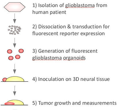
BlastomaBrain is a drug testing service to investigate the efficiency of compounds against brain tumors. It allows us to study glioblastomas in their natural environment, within healthy human neural tissue. Thus, responses to drugs are also likely to be closer to reality.
We transduce human glioblastoma cells to overexpress a fluorescent reporter and generate tumor spheroids. These are inoculated on our 3-dimensional neural tissue derived from human pluripotent stem cells. Tumor growth is monitored through the measurement of the fluorescent signal.
Long-term compound testing, 3D human organoids, as well as realistic physiological neural-tumoral interactions make the BlastomaBrain advantageous when compared to current 2D and 3D tumor cells, and animal models.
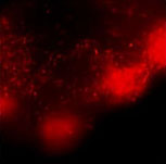
GLIOBLASTOMA SPREADS AMONG NEURAL TISSUE
The fluorescent glioblastoma grows rapidly inside the neural tissue. Semi-automatic quantification is applied to measure tumor area, in parallel, we monitor tumor dispersion and metastasis.
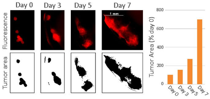
Fig. A: Topview of glioblastoma growth in the neural tissue 0-7 days post-inoculation. Fluorescence tumor imaging on the upper panel is quantified in a semi-automatized manner to obtain glioblastoma area in the bottom and right panels.
SEMI-AUTOMATIC QUANTIFICATION AND HISTOLOGICAL READOUTS
Semi-automatic fluorescence quantification revealed that the anti-cancer drug Temozolomide strongly affects glioblastoma growth. Our absolute IC50 of 258uM is comparable to the literature data. Alternative histologic, proteomic, or genomic readouts can also be performed.
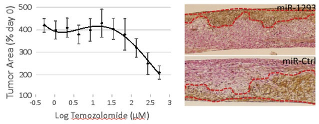
Fig. B: The left panel shows semi-automatized quantification of tumor fluorescence area following Temozolomide treatment for 7 days (3 replicates). An alternative histological readout can be performed to visualize tumor growth which was altered by miR-1293 micro-RNA on the right panel (Vimentin IHC, braun colored).
BLASTOMABRAINTM GLIOBLASTOMA INVASION MODEL - SERVICE SPECIFICATIONS
|
Cell types |
Tumor: Spheroids from continuous and patient-derived human glioblastoma cells, inoculated on the 3D neural tissue described below. 3D Neural tissue: MinibrainTM generated from human pluripotent stem cells. Composed of neurons, astrocytes, oligodendrocytes, and neural progenitor cells. The latter cells keep generating newborn neurons in a dynamic process. |
|
Production technology |
Neurix’s MinibrainTM & Neurosphere technology with minimum batch-to-batch variability guaranteed by extensive quality control of identity (rt-qPCR) |
|
Field of application |
Lead compound validation |
|
Assay window |
Short-term (7 days) to long-term (one month) |
|
Readouts |
- Tumor area (growth and motility) - Total fluorescence (growth) - Maximum radius (motility) - Number of secondary loci/metastasis (motility) - Number of secondary loci/metastasis (motility) |
OUR PUBLICATIONS
Cosset, E. et al. Human tissue engineering allows the identification of active miRNA regulators of glioblastoma aggressiveness. Biomaterials 107, 74–87 (2016).
Nayernia, Z. et al. The relationship between brain tumor cell invasion of engineered neural tissues and in vivo features of glioblastoma. Biomaterials 34, 8279–8290 (2013).
VALIDATED ASSAYS AND PROTOCOLS
The BlastomaBrain service seamlessly integrates with a wide range of validated assays, offering an invaluable resource for drug development and evaluating the efficacy of novel compounds.:
- Cell viability assays
- Histological analysis (IHC & IF)
- Cell sorting and cell population analysis (FACS)
- Genomic analysis
- Proteomic analysis
CONTACT US
At Neurix, we provide tailored services for various neural applications, such as gene/cell/polymer therapy testing, brain tumor drug testing, modeling neurodegenerative diseases, and conducting neurotoxicity assays. Our team of experienced scientists is dedicated to collaborating with you to comprehend your requirements and achieve your goals.
Email: info@neurix.ch
Phone: +41 22 379 46 43
Location:
Avenue de la Roseraie 64
1205 Geneva,
Switzerland